Quartz Imaging in Published Research
September 23, 2026
Most of Life on Earth Has Never Been Sequenced. Here Is One Piece Filled In.
Of all the fungal species estimated to exist, only about three to five percent have ever been formally described. The rest, millions of species, are what scientists call microbial dark matter: real, everywhere, and almost entirely unread, largely because they cannot be grown in a laboratory.
One of them lives somewhere unexpected: inside the gut of a winter stonefly.
Ejectosporus trisporus is a fungus that survives only in the hindgut of its insect host. Take it out, and it will not grow on any culture medium science has, which for decades made its genome effectively unreadable, since sequencing traditionally demands more clean DNA than a tiny gut symbiont can provide.
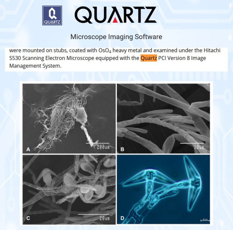
A team at the University of Toronto Scarborough found a way around that. Working from a single cluster of fungal threads picked out of a stonefly collected in Rouge National Urban Park, they amplified the trace of DNA present and assembled the first genome ever produced for this unculturable species, a quality on par with genomes built the traditional way from lab cultures.
To document the organism itself, they turned to the microscope. Scanning electron microscopy, captured on a Hitachi S530 using Quartz PCI, revealed the fine architecture of the fungal threads and both its asexual and sexual spores, while light microscopy showed the mature spores in detail. The two techniques together, one for ultrastructure, one for the living forms, built the visual record behind the genomic one.
Why chase a fungus most people will never see? Because these gut-dwelling fungi may be more sensitive indicators of freshwater quality than the insects that host them. Reading their genomes is a step toward understanding the hidden biology of the streams and rivers we depend on, and toward mapping the vast, mostly blank map of fungal life on Earth.
Every genome sequenced turns a little more dark matter into knowledge.
All rights for the excerpts and images remain with the authors and the full article is foundhere
Sriram, S., Alsafar, H., Lusa, R., & Wang, Y. (2026). Single-thallus genomics of Ejectosporus trisporus, an unculturable stonefly gut fungal symbiont. Environmental Microbiology.
Thank you for mentioning Quartz Imaging in the paper and congratulations on publishing!
Learn more about Quartz Imaging products.
September 16, 2026
Some Discoveries Take Ten Years, and a Little Help From Those Who Came Before.
Not every scientific discovery announces itself. Some are hiding in plain sight, waiting for someone patient enough to look closely.
While examining lichens across eastern North America, the late Richard Harris noticed something odd: a tiny parasitic fungus living on a common lichen, so small it is invisible to the naked eye and easily mistaken for part of the host. He began drafting a description of it in 2018. Then came delays, then the pandemic, and in 2021 Harris passed away before the work was finished.
His colleagues carried it forward. After examining more than 1,500 specimens across herbarium collections and years of fieldwork, they completed what he had started. This year, more than a decade after the project began, the fungus was formally named and described as a species new to science: Minutophoma buelliarum.
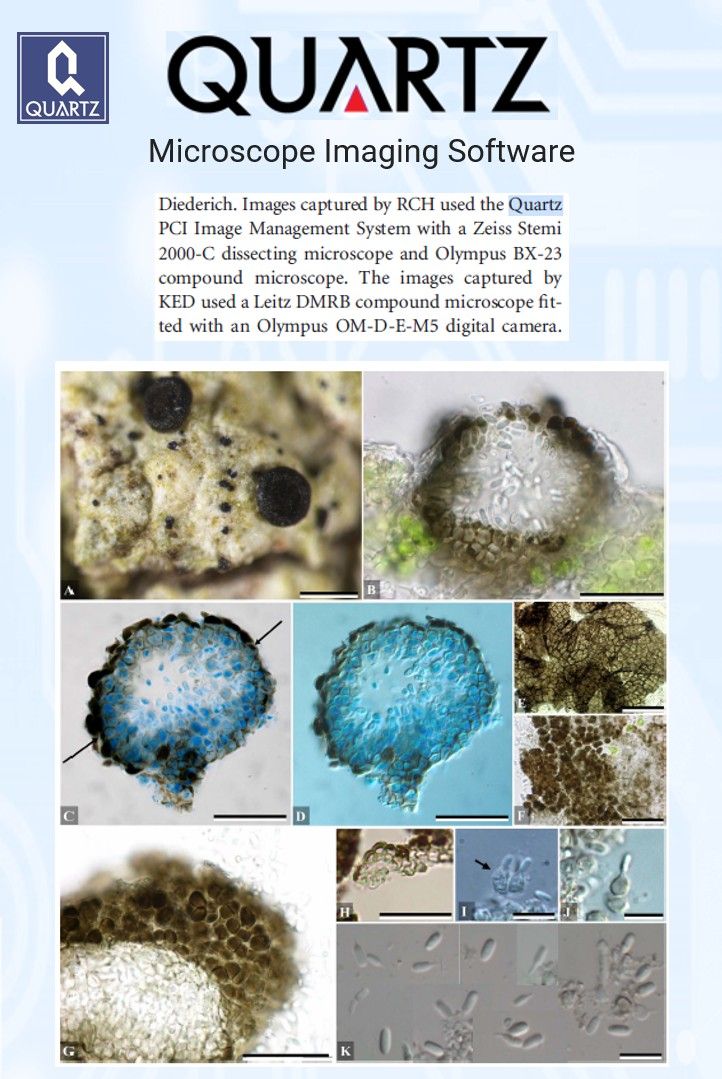
The work is a reminder of how much careful looking real discovery demands. To document a fungus this small, the team relied on light microscopy, capturing the fine architecture of its spore-producing structures through dissecting and compound microscopes. Those images, some acquired using Quartz PCI, made it possible to measure features just a few micrometres across and to distinguish this fungus from the several look-alikes sharing the same host.
It is easy to picture microscopy as the world of electron beams and nanometres. But a great deal of foundational science still happens at the eyepiece of a light microscope, where a trained eye and a good image separate one species from another. Naming the living world, one small organism at a time, is how we come to understand the biodiversity around us, including the quiet, overlooked players most people will never see.
This one carries an added meaning. It stands as a finished tribute to the researcher who first noticed it, and a reminder that science is a relay, carried forward by those who pick up where others left off.
All rights for the excerpts and images remain with the authors and the full article is foundhere
Driscoll, K. E., Lendemer, J. C., Clayden, S. R., & Harris, R. C. (2026). Minutophoma buelliarum (Ascomycota, subphylum Pezizomycotina), a new lichenicolous fungus widespread on corticolous, norstictic acid-producing Buellia in eastern North America. The Bryologist, 129(2), 130–139.
Thank you for mentioning Quartz Imaging in the paper and congratulations on publishing!
Learn more about Quartz Imaging products.
September 09, 2026
Building a Safer, Cheaper Battery From Earth-Abundant Materials.
The batteries powering our phones, cars, and power grids mostly rely on lithium. But lithium is unevenly distributed, increasingly expensive, and its flammable liquid electrolytes carry real safety risks. As the world electrifies, the search is on for a battery built from cheaper, safer, more abundant ingredients.
The Obstacle:
Zinc is a promising alternative. It is abundant, inexpensive, and works with water-based electrolytes, which makes it far safer than lithium's organic ones. The catch is the cathode, the electrode where the energy is stored. Vanadium oxide is a strong candidate, but on its own it conducts electricity poorly, degrades over repeated charging, and slowly dissolves into the electrolyte, cutting the battery's life short.
The Solution:
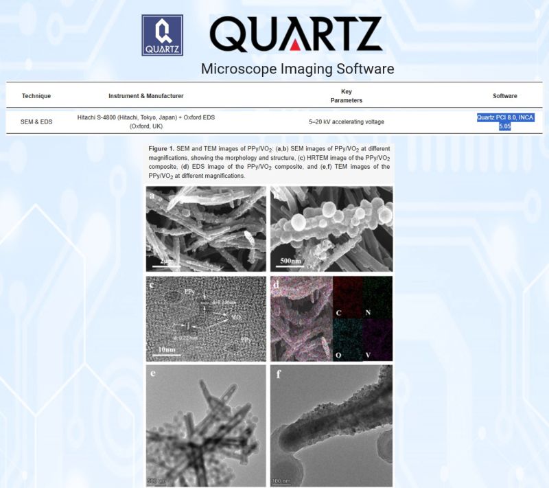
A research team at Hunan University of Humanities, Science and Technology tackled this by pairing vanadium oxide with polypyrrole, a flexible, electrically conductive polymer. They grew tiny vanadium oxide spheres directly onto polypyrrole nanorods, creating a core-and-shell structure where the polymer forms a conductive, protective scaffold around the active material. Scanning electron microscopy, with images acquired through Quartz PCI, confirmed the architecture, showing the nanospheres anchored along the rods.
The result:
The composite delivered more than double the capacity of vanadium oxide alone, and it held on to 87 percent of its capacity after 1,200 charge-discharge cycles. The conductive polymer network cut electrical resistance and shielded the active material from dissolving away.
The Humanity:
Better energy storage is one of the quiet foundations of a cleaner future. Solar panels and wind turbines only help if we can store what they generate, and safer, cheaper batteries made from abundant materials bring that closer for homes, grids, and communities. Progress toward clean energy often comes down to work like this, at the scale of a single engineered particle.
Sometimes the path to a bigger, cleaner grid runs through the smallest details of a material's design.
All rights for the excerpts and images remain with the authors and the full article is foundhere
Zhou, T., Li, S., Xie, D., Liu, Y., Cheng, Y., & Li, X. (2025). Hydrothermally synthesized PPy/VO2 nanorod composites for high-performance aqueous zinc-ion battery cathodes. Micromachines, 16(6), 705.
Thank you for mentioning Quartz Imaging in the paper and congratulations on publishing!
Learn more about Quartz Imaging products.
September 03, 2026
Locking Vitamins Into Food, One Tiny Crystal Network at a Time.
Vitamin E is one of those quiet essentials. It supports the immune system, protects our cells, and carries real antioxidant power. But it has a frustrating weakness: it is fragile. Exposed to light, heat, or oxygen, it breaks down quickly, which makes it hard to keep intact in the foods meant to deliver it.
The Obstacle:
Vitamin E dissolves in fat, so it naturally travels in the oily parts of food. The problem is that liquid oil offers little protection, and the vitamin degrades before it can do its job. Food scientists needed a way to hold both the oil and the vitamin in a stable structure, without resorting to the hydrogenated or saturated fats the industry is trying to move away from.
The Solution:
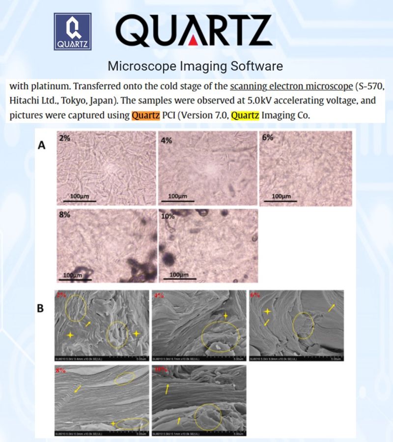
A team at Jiangsu University turned to oleogels, liquid oils trapped inside a solid-like network built from tiny edible crystals. Using a food-safe structuring agent called monostearin, they built gels around vitamin E and soybean oil, then tested how well the structure held together and shielded the vitamin. To see the network itself, they used cryo-scanning electron microscopy, with the images captured through Quartz PCI, revealing the delicate needle-like crystals and stacked lamellar layers that lock the oil in place.
The result:
The more crystal network the gel contained, the better it performed. The denser gels held their oil more tightly and, crucially, protected the vitamin far better under UV light. After fifteen days of light exposure, plain oil had lost nearly half its vitamin E, while the strongest gels retained the vast majority of it.
The Humanity:
This is a quiet but practical step toward healthier food. An edible gel that guards fragile nutrients and replaces less healthy fats could help fortify everyday products, from baked goods to functional foods, and deliver vitamins that actually survive to the plate. Small structures, engineered with care, doing real nutritional work.
Sometimes protecting what matters comes down to building the right structure at the smallest scale, and making it visible.
All rights for the excerpts and images remain with the authors and the full article is foundhere
Monto, A. R., Yuan, L., Xiong, Z., Shi, T., Li, M., Wang, X., Jin, W., Li, J., & Gao, R. Physicochemical properties of α-tocopherol and soybean oil-loaded oleogel developed with glyceryl monostearate.
Thank you for mentioning Quartz Imaging in the paper and congratulations on publishing!
Learn more about Quartz Imaging products.
August 19, 2026
Before a Water Filter Can Save Lives, It Has to Survive Getting Wet.
It sounds obvious.
But almost no one had actually tested it.
Polymer inclusion membranes are a promising, low-cost way to pull contaminants like arsenic out of drinking water. They work by trapping an ionic liquid carrier inside a polymer film, which grabs the pollutant as water passes through.
The Obstacle:
These membranes are designed to sit in water for hours or days at a time.
Yet almost no research had asked a basic question: what does prolonged water contact actually do to the membrane itself?
If the active ingredient washes away, the membrane can fail exactly when it is needed most.
The Solution:
A research team at the University of Girona in Spain set out to find out.
They built membranes from two common polymers, cellulose triacetate and PVC, each loaded with different versions of the ionic liquid Aliquat 336.
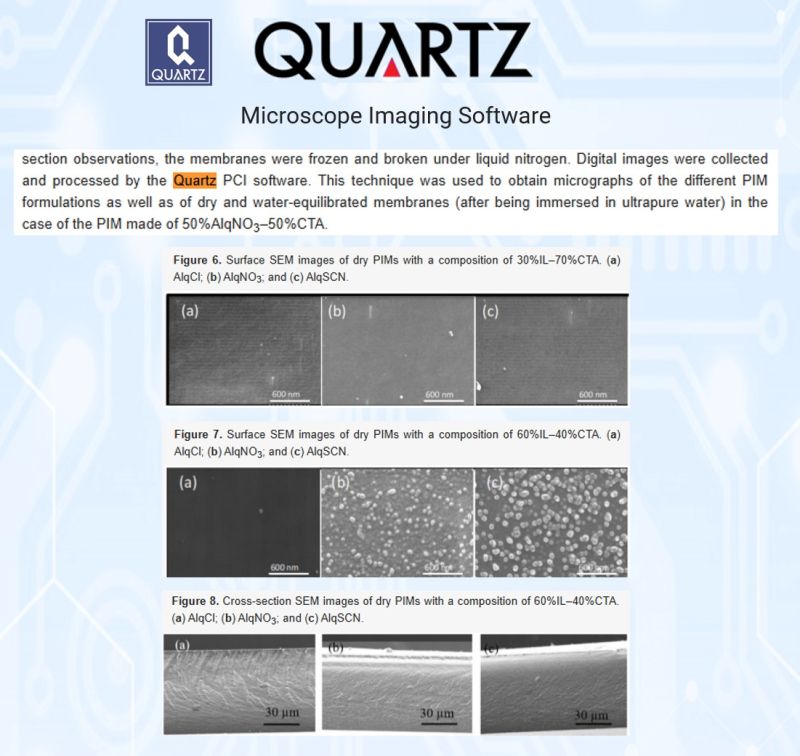
Using a Hitachi FE-SEM microscope with Quartz PCI software to capture and process the images, they compared the surface of each membrane before and after four days submerged in ultrapure water.
The imaging told a clear story. Some membranes showed nano-sized clusters of ionic liquid on their dry surface that disappeared entirely after water contact, evidence of real material loss, even though the bulk of the membrane underneath stayed intact.
The Humanity:
The team then tested the most water-stable version on real arsenic removal, and it held up.
The membrane kept working at roughly 79 percent efficiency across five full reuse cycles.
That matters far beyond the lab. Arsenic contamination in drinking water affects hundreds of millions of people worldwide, many in places where an affordable, reusable filter is the only realistic option. Knowing exactly how these membranes age in water is what turns a promising material into one that communities can actually depend on.
All rights for the excerpts and images remain with the authors and the full article is foundhere
Fontàs, C., Vera, R., Anticó, E., Martínez de Yuso, M.d.V., Rodríguez-Castellón, E., & Benavente, J. (2022). New Insights on the Effects of Water on Polymer Inclusion Membranes Containing Aliquat 336 Derivatives as Carriers. Membranes, 12(2), 192.
Thank you for mentioning Quartz Imaging in the paper and congratulations on publishing!
Learn more about Quartz Imaging products.
August 12, 2026
Unraveling a 160-Year-Old Puzzle in Parasitic Plant Identification
Cuscuta, commonly known as dodder, is a leafless parasitic plant with no roots or foliage to rely on for identification.
That leaves flowers, fruit, and inflorescences as the primary tools for telling its roughly 200 species apart.
Researchers have used inflorescence structure in Cuscuta species descriptions since 1859. Yet until recently, no comprehensive study had ever mapped out that diversity across the genus.
The Obstacle:
Cuscuta inflorescences are notoriously difficult to classify. Historical descriptions borrowed racemose terms like "umbelliform" or "paniculiform" to approximate branching patterns that are, in reality, monochasial cymes. The result was decades of inconsistent terminology.
The Solution:
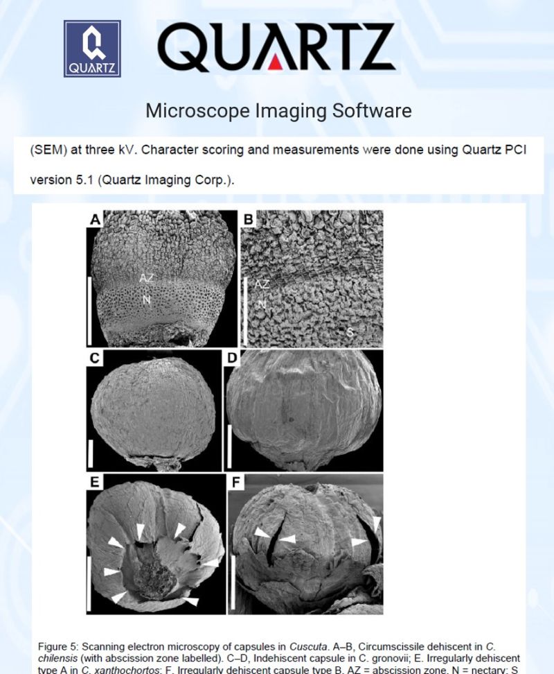
A research team at Wilfrid Laurier University examined 132 Cuscuta taxa using herbarium specimens, and grew eight living species to study inflorescence development firsthand. Using scanning electron microscopy, the team relied on Quartz PCI for character scoring and precise measurements of inflorescence structures.
This work identified three major architectural types across the genus: "Monogynella," "Cuscuta," and "Grammica," each tracing back to a shared fundamental unit, the scorpioid monochasial cyme.
The Humanity:
Several Cuscuta species are aggressive agricultural weeds, capable of devastating crops and persisting in soil for decades. Clearer identification tools support better biodiversity management and more effective control strategies for growers and researchers alike.
Sometimes the smallest structural details carry the biggest implications for how we manage the natural world around us.
All rights for the excerpts and images remain with the authors and the full article is foundhere
Glofcheskie, Morgan, "Diversity and Evolution of Inflorescences in Cuscuta" (2022). Theses and Dissertations (Comprehensive). 2502.
Thank you for mentioning Quartz Imaging in the paper and congratulations on publishing!
Learn more about Quartz Imaging products.
July 29, 2026
When Growing New Tissue, the Scaffold Is Everything. So How Do You Build It on Purpose?
Tissue engineering holds a remarkable promise: growing replacement tissue in the lab, on tiny scaffolds that give cells a structure to attach to and organize around. But those scaffolds only work if their architecture is right. Too dense and cells cannot infiltrate; too sparse and the structure collapses. Getting it right has long been a matter of trial and error.
The Obstacle:
One of the best ways to make these scaffolds is melt electrospinning, which uses an electric field to draw molten biodegradable polymer into fine fibers that stack into a sponge-like mesh. The trouble is control. Fiber thickness and porosity depend on a tangle of interacting settings, and researchers have largely had to guess, run the machine, image the result, and try again. That is slow, and it wastes material.
The Solution:
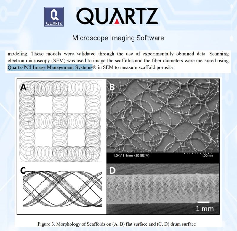
A team at the University of Victoria, here in British Columbia, set out to replace the guesswork with prediction. They built a mathematical model linking the key machine settings, nozzle size, applied voltage, and the distance to the collector, to the two things that matter most in the finished scaffold: fiber diameter and porosity. Then they tested it against real scaffolds made from PCL, a common biodegradable polymer.
To validate the model, the team imaged the scaffolds by scanning electron microscopy on a Hitachi S-4800 and used Quartz PCI to measure the fiber diameters, loop dimensions, and overlap that define the mesh. Those measurements were the proof: the model's predictions of fiber size and porosity matched the fabricated scaffolds closely, within a few percent across a wide range of settings.
The Humanity:
Turning scaffold fabrication from trial and error into something a researcher can dial in on purpose is a quiet but meaningful step. It means less wasted material, faster iteration, and scaffolds engineered to a specification rather than stumbled upon. For the long road toward lab-grown tissue for wound healing, vascular grafts, and regenerative medicine, predictability is what lets the field move from art to engineering.
Sometimes progress is not a new discovery, but the ability to make the same thing reliably, every time.
All rights for the excerpts and images remain with the authors and the full article is foundhere
Ko, J., Bhullar, S. K., Mohtaram, N. K., Willerth, S. M., & Jun, M. B. G. (2014). Using mathematical modeling to control topographical properties of poly (ε-caprolactone) melt electrospun scaffolds. Journal of Micromechanics and Microengineering, 24(6), 065009.
Thank you for mentioning Quartz Imaging in the paper and congratulations on publishing!
Learn more about Quartz Imaging products.
July 15, 2026
When You Have More Juice Than You Can Sell, What Do You Do?
For lemon and grapefruit growers, seasonality is both a blessing and a curse. When harvests peak, prices crash. When supply dries up, growers miss the market entirely. For decades, the answer has been to accept the loss. A new approach asks a different question: what if we could preserve the harvest and add value at the same time?
The Obstacle:
Fruit juices have short shelf lives. Fresh lemon juice, rich in nutrition and flavor, turns within days. Dehydrating it into a stable powder should work, but commercial encapsulating agents—the coatings that protect juice particles during the drying process—are expensive, scarce, and often come from far away. Gum arabic, the traditional choice, costs more than many farmers can justify.
The Solution:
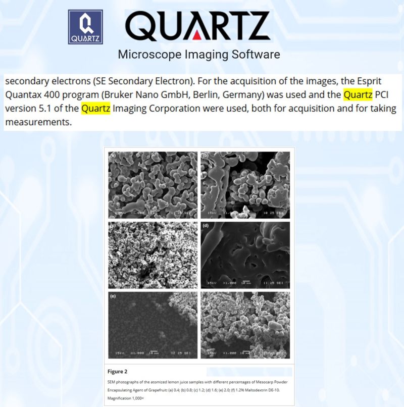
Researchers at Murcia, ISA, and Florida International Universities explored whether a waste product from grapefruit processing could work as well. They spray-dried lemon juice with varying concentrations of powdered grapefruit mesocarp, the fibrous material normally discarded. Using scanning electron microscopy to observe the resulting particles, they measured size and morphology with precision using Quartz PCI to understand how the encapsulant performed.
The result:
at 1.2% concentration, the grapefruit mesocarp produced stable, spherical particles between 3 and 6 micrometers in size. The glass transition temperature remained stable across all treatments, indicating good shelf stability. And the encapsulant came from agricultural waste.
The Humanity:
One waste stream becomes a solution for another. Grapefruit processors gain a value-added product to sell. Lemon growers can preserve their harvest at peak season, stabilizing income and reducing waste. Consumers get access to a nutritious product year-round without the chemical complexity of synthetic encapsulants. A byproduct becomes a resource.
In food processing, as in nature, waste is often just a resource waiting for the right use.
All rights for the excerpts and images remain with the authors and the full article is found here
Alcantara-González, C.S., Escotto-Díaz, M.A., García, E., Rodríguez-Jiménez, J.J. (2019). Spray drying of lemon juice using pulverized grapefruit mesocarp as encapsulating agent. Food Science & Nutrition, 7(10), 3325–3333.
Thank you for mentioning Quartz Imaging in the paper and congratulations on publishing!
Learn more about Quartz Imaging products.
June 24, 2026
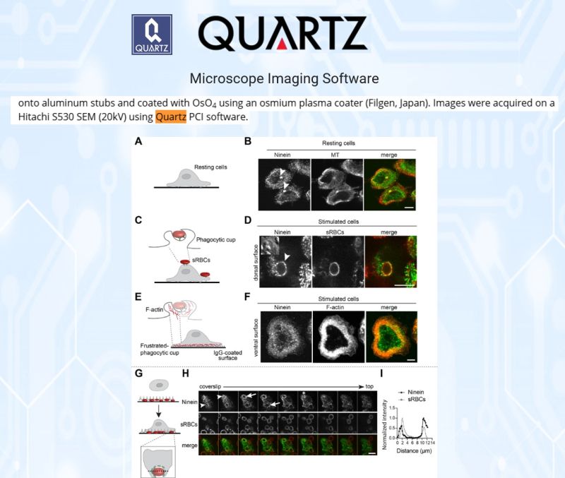
How Your Immune Cells Decide What to Swallow, and What It Could Teach Us About Disease.
Every day, your immune cells engulf threats, dead cells, debris, and invading microbes, in a process called phagocytosis. It is one of the oldest defences we have. Yet exactly how a macrophage reaches out and pulls a target inward is still only partly understood.
The Obstacle:
Phagocytosis is driven by F-actin, the protein scaffolding that lets a cell wrap membrane arms around a particle. But the role of microtubules, the cell's other structural network, and the proteins guiding them, has been far less clear.
The Solution:
Researchers at the University of Toronto Scarborough, publishing in Molecular Biology of the Cell, studied ninein, a protein known for organising microtubules, to learn what it does during phagocytosis.
To capture the surface architecture of the process, the team used scanning electron microscopy on a Hitachi S530, with images acquired through Quartz PCI software. The micrographs made the defect strikingly visible: where healthy cells wrapped membrane arms tightly around their targets, ninein-depleted cells left many particles bare.
The result:
Ninein is needed right at the first step, the moment of particle engagement, where it drives early F-actin recruitment and forms the cup that begins engulfment. It also helps assemble the dynein motor machinery that draws the captured particle inward.
The Humanity:
Phagocytosis sits at the centre of how we fight infection, clear dying cells, and regulate inflammation. When it falters, the effects reach into immune deficiency, chronic inflammation, and how tumours evade the immune system. Mapping the proteins that govern the first seconds of engulfment is how researchers learn to one day tune that response.
The smallest movements inside a single cell, made visible, are where that understanding begins.
All rights for the excerpts and images remain with the authors and the full article is found here
Omer, S., Li, J., Yang, C. X., & Harrison, R. E. (2024). Ninein promotes F-actin cup formation and inward phagosome movement during phagocytosis in macrophages. Molecular Biology of the Cell.
Thank you for mentioning Quartz Imaging in the paper and congratulations on publishing!
Learn more about Quartz Imaging products.
June 17, 2026
A New Species Was Just Discovered — Hidden Inside a Vulnerable Seagrass in Southeast Asia.
Seagrasses are among the most important ecosystems on the planet.
They shelter juvenile fish, sequester carbon, and stabilize coastlines. Yet the microscopic threats living inside them remain largely uncharted.
A research team led by Viktorie Kolátková at the University of Victoria set out to change that.
The Obstacle:
The genus Marinomyxa consists of obligate intracellular parasitic protists known to infect tropical seagrasses of the genus Halophila. Until now, only two species had been formally described. But mounting evidence suggested the diversity of these parasites was far greater than morphology alone could reveal.
The Solution:
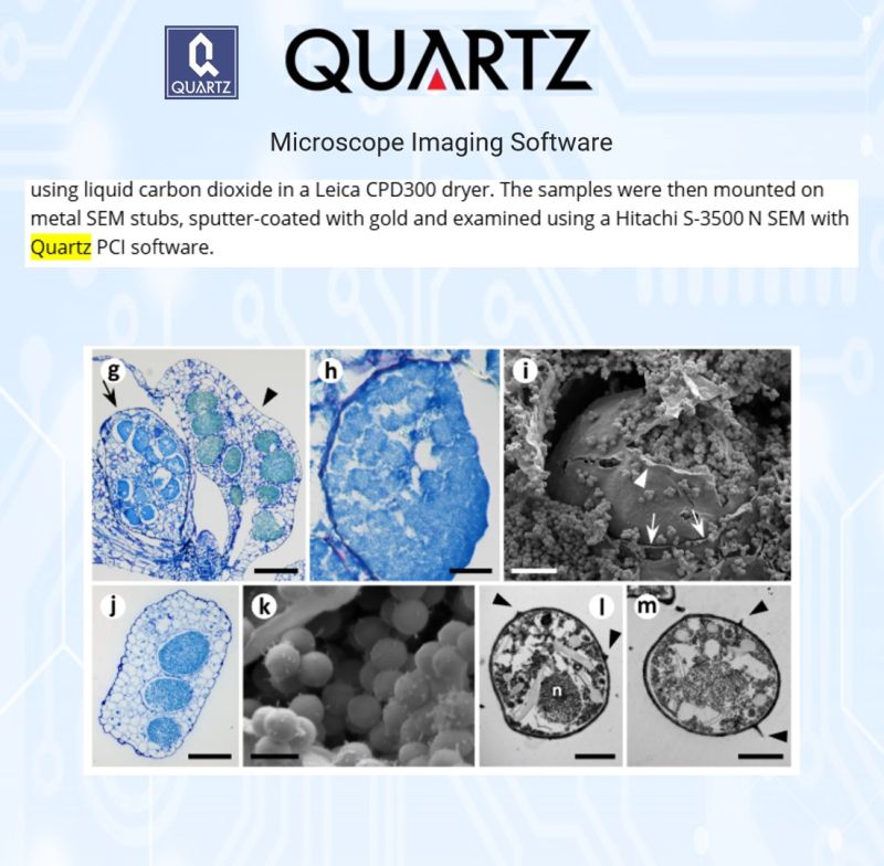
The team combined molecular sequencing, phylogenetic analysis, and microscopy to investigate Marinomyxa infections across multiple Halophila species and geographies, from Thailand and the Philippines to Puerto Rico and New Caledonia. Scanning electron microscopy was performed using a Hitachi S-3500N SEM, with Quartz PCI software used for image acquisition. The SEM micrographs provided critical visual confirmation of the characteristic cell-wall protuberances on resting spores, a defining feature used to identify and differentiate Marinomyxa species.
The findings revealed two significant discoveries. First, a previously undescribed parasite in the vulnerable intertidal seagrass Halophila beccarii from Southeast Asia, now formally named Marinomyxa denhartogi sp. nov. in honour of the late Dutch botanist Cornelis den Hartog, who first observed the organism in 1996. Second, the cosmopolitan seagrass Halophila decipiens from Puerto Rico was confirmed as an additional host of Marinomyxa halophilae, extending the known range of this parasite well beyond the Indo-Pacific.
The Humanity:
Seagrass ecosystems are under mounting pressure from climate change, biological invasions, and coastal development. Understanding the parasites that live within them is not an academic exercise. It is a prerequisite for predicting how these ecosystems will respond as conditions change, and for protecting the communities and species that depend on them.
All rights for the excerpts and images remain with the authors and the full article is found here
Kolátková, V., Rattanachot, E., Dieppa, A., & Gawryluk, R.M.R. (2026). First report of Marinomyxa spp. (Rhizaria: Endomyxa: Phytomyxea) infection in seagrasses Halophila beccarii and Halophila decipiens: Supporting evidence for a long-standing symbiosis. Plant Pathology.
Thank you for mentioning Quartz Imaging in the paper and congratulations on publishing!
Learn more about Quartz Imaging products here.
June 10, 2026
Healthier Animals, Fewer Antibiotics. Science Is Showing Us How.
Weaning is one of the most stressful transitions a young animal faces.
The rumen is still developing. The immune system is under pressure. And in commercial livestock production, the traditional response has been antibiotics, which work, but at a cost to the animal's microbial balance and long-term health.
Researchers at the Gansu Academy of Agricultural Sciences and Gansu Agricultural University set out to find a better path.
The Obstacle:
Young Hu sheep transitioning off maternal milk need support for rumen development and immune function. The question was whether a natural probiotic could deliver that support reliably, at the right dose, over time.
The Solution:
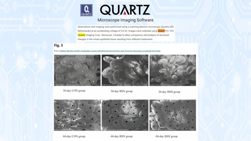
The team fed 45 weaned male lambs one of three diets for 60 days: a standard basal diet, or the same diet supplemented with Bacillus subtilis at either 100 mg/kg or 200 mg/kg. Rumen tissue was collected at 30 and 60 days and examined using a FEI Quanta 200 scanning electron microscope. Quartz PCI 10.0 was used to acquire and analyze the SEM images, capturing structural changes in the rumen epithelial tissue across treatment groups.
The Humanity:
The results were clear. Both supplementation groups showed taller rumen papillae, higher total volatile fatty acid production, and reduced inflammatory markers compared to the control.
After 60 days, the 100 mg/kg dose proved optimal, sustaining the structural and immune benefits without the microbiome disruption seen at higher doses.
Better rumen development means more efficient feed conversion. A stronger immune system means healthier animals with less reliance on antibiotics. That matters for farmers, for food systems, and for the animals themselves.
All rights for the excerpts and images remain with the authors and the full article is found here
Zhao, H., Liu, L., Wang, Z., Sha, Y., & Song, S. (2026). Dietary Bacillus subtilis modulates rumen epithelial barrier function and immune responses in weaned Hu sheep. Scientific Reports, 16, 17089.
Thank you for mentioning Quartz Imaging in the paper and congratulations on publishing!
Learn more about Quartz Imaging products here.
June 03, 2026
Protecting the Strawberry Fields of British Columbia: One Tiny Weevil at a Time.
A destructive pest arrived in southwestern British Columbia in 2019.
The strawberry blossom weevil (Anthonomus rubi) cuts flower buds from their stems before they can bloom, causing serious damage to strawberry and other berry crops in the Rosaceae family.
The problem:
It arrived without the natural enemies that keep its populations in check in Europe, its native range.
That is the obstacle growers and researchers are still working to solve.
Jade Sherwood, through the University of British Columbia's Faculty of Graduate and Postdoctoral Studies, took a systematic approach.
Working with collaborators from the EU-based Centre for Agriculture and Biosciences International (CABI), the research surveyed the weevil across 12 European countries to identify parasitoid species that naturally control it.
The result:
12 parasitoid species identified, including seven not previously recorded as being associated with A. rubi.
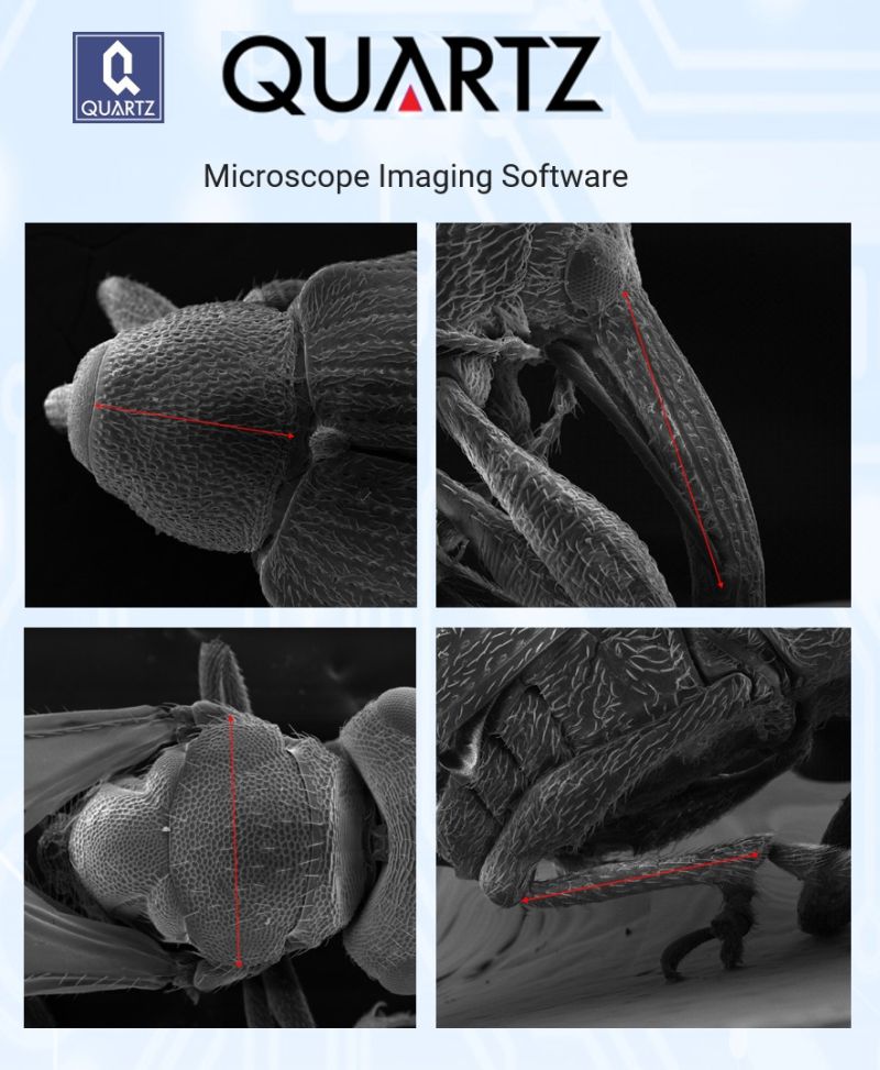
To document the weevil's morphology with precision, the team used a Hitachi S-2500 scanning electron microscope, with Quartz PCI software handling image acquisition, measurement, and annotation.
SEM images were captured at magnifications of 70x to 110x at 10kV, producing the high-resolution reference data needed to accurately measure and compare specimens across pronotum, rostrum, and tibia structures.
In the introduced range of BC, the research also found that larger strawberry buds produce larger, likely fitter weevils, a finding with direct practical implications for how future biocontrol agents should be reared and deployed.
This is the groundwork that makes targeted, chemical-free pest management possible: protecting the berry crops that farmers and communities in BC depend on.
All rights for the excerpts and images remain with the authors and the full thesis is found here
Sherwood, J. (2025).
Native range surveys for natural enemies of Anthonomus rubi and effects of host plant bud size in the introduced range [Master's thesis, University of British Columbia]. UBC cIRcle.
Thank you for mentioning Quartz Imaging in the thesis and congratulations on completing your Master of Science, Jade!
Learn more about Quartz Imaging products here.
May 27, 2026
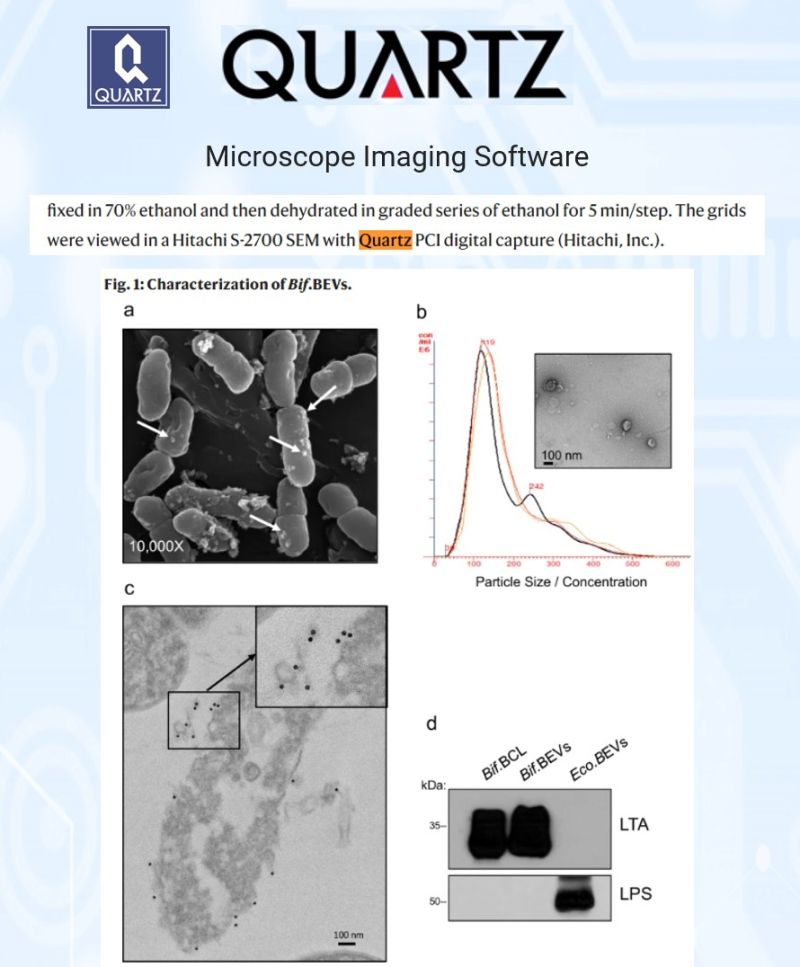
What if the key to unlocking cancer immunotherapy was already living inside you?
Lung cancer remains the leading cause of cancer-related deaths worldwide. Immunotherapy has changed the game, but too many patients still do not respond. Scientists have long suspected gut bacteria play a role. Now, there is evidence of exactly how.
A research team publishing in Nature Communications identified that tiny nano-sized vesicles shed by Bifidobacterium, a common gut commensal, can travel from the intestine, cross tissue barriers, and accumulate directly inside lung tumors.
Once there, they prime tumor cells to express more PD-L1. That makes the cancer more visible, and more vulnerable, to anti-PD-1 immunotherapy.
In mouse models, combining these vesicles with anti-PD-1 significantly reduced tumor growth, increased tumor-infiltrating CD8+ T cells, and shifted the tumor immune environment toward an anti-cancer state.
To map exactly where and how this was happening inside tumor tissue, the team turned to spatial profiling using the NanoString GeoMx Digital Spatial Profiling instrument, with image analysis supported by Quartz PCI.
The result: a measurable, spatially resolved picture of PD-L1 upregulation and immune cell infiltration across treatment groups.
The gut-lung axis is no longer a hypothesis. It is a potential therapeutic target, and imaging made it visible.
All rights for the excerpts and images remain with the authors and the full article is found here.
Preet, R., Islam, M.A., Shim, J. et al. Gut commensal Bifidobacterium-derived extracellular vesicles modulate the therapeutic effects of anti-PD-1 in lung cancer. Nature Communications 16, 3500 (2025).
Thank you for mentioning Quartz Imaging in the paper and congratulations on publishing!
Learn more about Quartz Imaging products here.
May 20, 2026
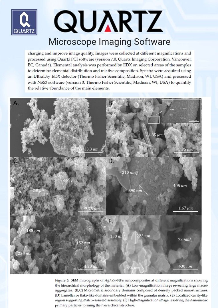
Clean water is not a luxury. It is a right. And science is finding new ways to protect it.
Synthetic dyes from industrial processes are among the most persistent and harmful contaminants in our water systems.
Conventional treatment methods struggle to break them down.
A research team at the University of Cagliari set out to change that. They developed nanocomposites using a simple, sustainable ingredient: an extract from the microalgae Chlorella vulgaris.
No hazardous chemicals. No energy-intensive processes.
The result was a photocatalytic material capable of breaking down Congo Red, a toxic industrial dye, using UV light.
To characterize the material at the nanoscale, the team used Quartz PCI to process their SEM micrographs, capturing the hierarchical structure of the nanocomposites with precision.
The findings: up to 49% dye removal in 180 minutes, at a low catalyst loading, with performance that held steady across varying light intensities.
That last point matters. It means the system does not need to be pushed harder to work. It is already operating efficiently.
Cleaner water, greener chemistry, and a more rational use of energy. That is the direction this research points.
All rights for the excerpts and images remain with the authors and the full article is found here.
Zedda, F., Atzori, F., Casu, S., Sidorowicz, A., Fais, G., Desogus, F., Licheri, R., Porcu, S., Cao, G., Lutzu, G.A., & Concas, A. (2026). Green-Synthesized Ag/Zn Nanocomposites from Chlorella vulgaris Polar Extract: Sustainable Photocatalytic Water Remediation and Kinetic Modeling. Sustainability, 18, 4607.
Thank you for mentioning Quartz Imaging in the paper and congratulations on publishing!
Learn more about Quartz Imaging products here.
May 16, 2026
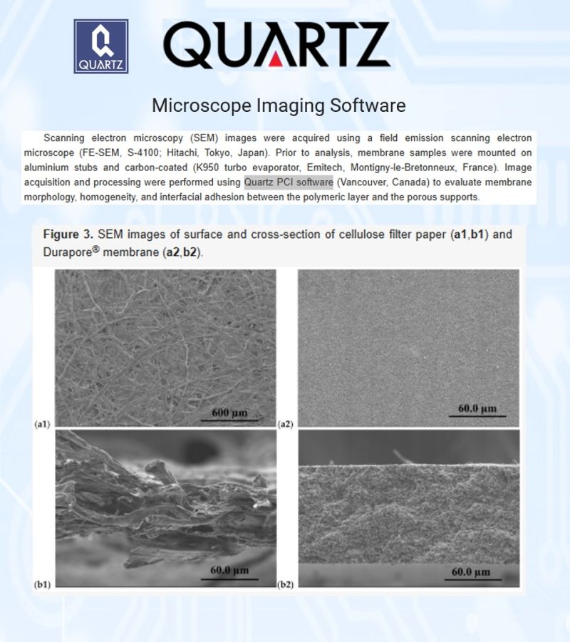
Cleaner Water for More People Could Start with Using Less to Make More
Arsenic in drinking water is a global health crisis affecting hundreds of millions of people.
Existing filtration membranes can remove it, but they are expensive and material-intensive to produce.
A research team at the University of Girona in Spain set out to change that.
Their approach was to deposit polymer inclusion membranes onto simple porous supports, such as cellulose filter paper and commercial PVDF membrane, using up to 75% less material than conventional methods.
To confirm the membranes were properly formed and structurally sound, the team used scanning electron microscopy with Quartz PCI software to evaluate surface morphology, layer uniformity, and the adhesion between the polymer phase and the porous support.
The results were compelling. The supported membranes achieved 90 to 95% arsenic transport efficiency, comparable to standard membranes, and retained around 70% efficiency after three reuse cycles.
Less material. Equal performance. Reusable.
This is the kind of innovation that makes water purification technology more accessible, more sustainable, and ultimately more equitable for communities that need it most.
All rights for the excerpts and images remain with the authors and the full article is found here.
Khatir N, Anticó E, Fontàs C. A Novel Supported Polymer Inclusion Membrane Concept for Reagent-Efficient Membrane Design. Membranes. 2026;16(4):135.
Thank you for mentioning Quartz Imaging in the paper and congratulations on publishing!
Learn more about Quartz Imaging products here.
May 06, 2026
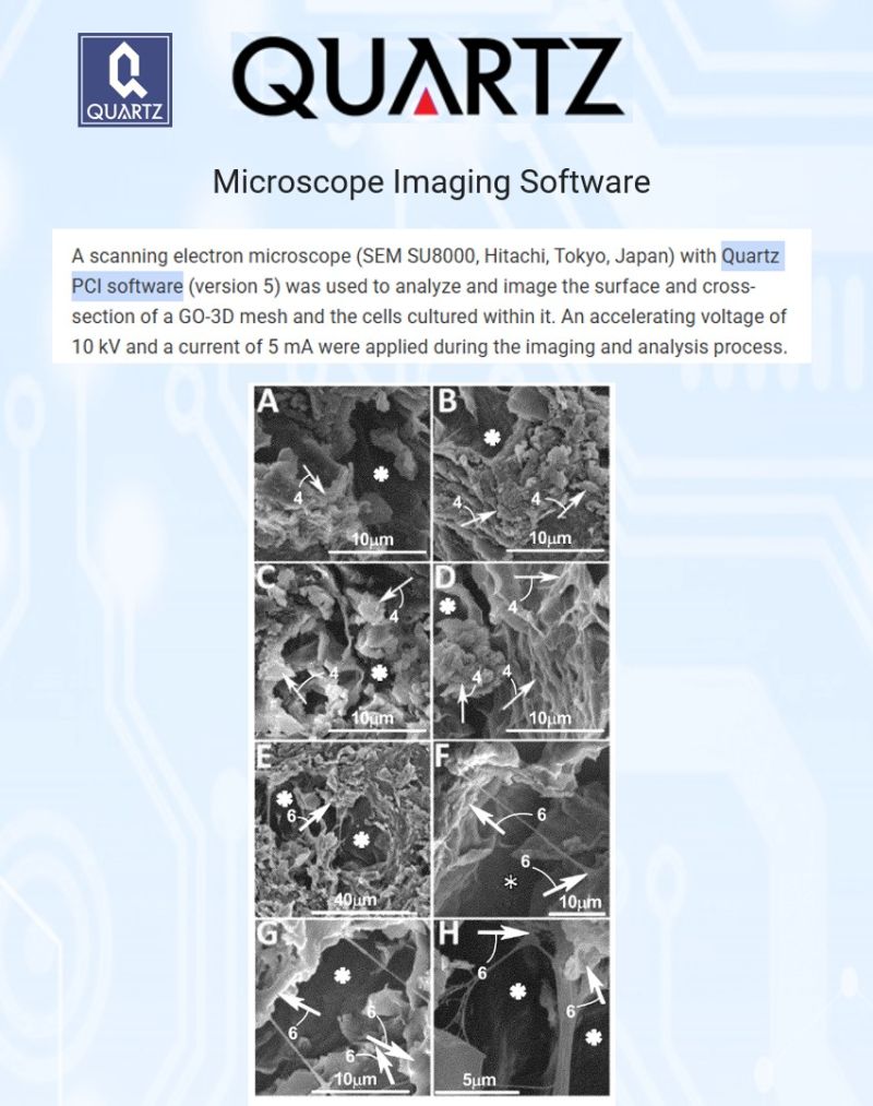
A 3D scaffold that could change how we grow cells — and one day, how we heal.
Traditional cell culture substrates have a fundamental limitation.
They are flat. Rigid. Poor at mimicking the real environment inside the human body.
Researchers at the University of North Dakota set out to change that.
The Obstacle:
Growing vascular cells in a way that reflects how they actually behave in living tissue requires a scaffold with tunable porosity, flexibility, and biocompatibility. Existing materials could not reliably deliver all three.
The Solution:
The team engineered a graphene oxide-based porous 3D mesh, combining graphene oxide with polyethylene glycol and a salt-leaching process to create a scaffold with precisely controllable pore size and mechanical properties. To characterize the mesh structure and analyze the cells cultured within it, the team used a scanning electron microscope paired with Quartz PCI software for imaging and pore size analysis across 500 pores per sample.
The Humanity:
When vascular endothelial cells and fibroblasts were cultured on this scaffold, they did not just survive. They organized themselves into structures resembling capillaries, the smallest blood vessels in the body. That is a meaningful step toward building lab environments that could accelerate drug testing, reduce animal trials, and eventually support tissue engineering for human medicine.
A patent granted. A foundation laid.
All rights for the excerpts and images remain with the inventors and assignee. The full patent is found here.
US Patent No. 12,077,776 B2 — Zhang, Y., Zhao, J.X., Darland, D. — University of North Dakota. Granted: September 3, 2024.
Congratulations to the inventors on the grant of this patent, and thank you for including Quartz PCI in your work.
Learn more about Quartz Imaging products here.
April 29, 2026
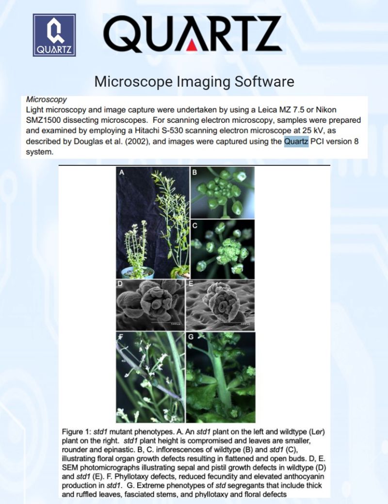
Understanding How Plants Grow Could Help Us Feed the World.
Every crop plant depends on healthy cell division and reproduction to thrive.
When those processes go wrong, yields drop, flowers fail, and growth stalls.
Understanding why is one of the most important questions in plant biology.
A research team at the University of Toronto, Scarborough set out to investigate what happens when a gene called HOP2, normally active only during meiosis, becomes overexpressed in the common research plant Arabidopsis.
The results were striking. Plants with elevated HOP2 levels showed stunted growth, misshapen flowers, altered leaf patterns, and severely reduced fertility.
To document the structural defects at the cellular level, the team used scanning electron microscopy with images captured using Quartz PCI.
The SEM micrographs revealed detailed floral organ abnormalities that would have been impossible to characterize with light microscopy alone.
The findings open a new window into how a single gene, when expressed at the wrong time or in the wrong place, can cascade into widespread developmental failure.
That knowledge brings us closer to understanding the genetic switches that govern plant health, resilience, and ultimately, food production.
All rights for the excerpts and images remain with the authors and the full article is found here.
Garrido AN, Francom T, Divan S, Kesserwan M, Daradur J, Riggs CD. Overexpression of HOP2 induces developmental defects and compromises growth in Arabidopsis. bioRxiv. 2021.
Thank you for mentioning Quartz Imaging in the paper and congratulations on publishing!
Learn more about Quartz Imaging products here.
April 22, 2026
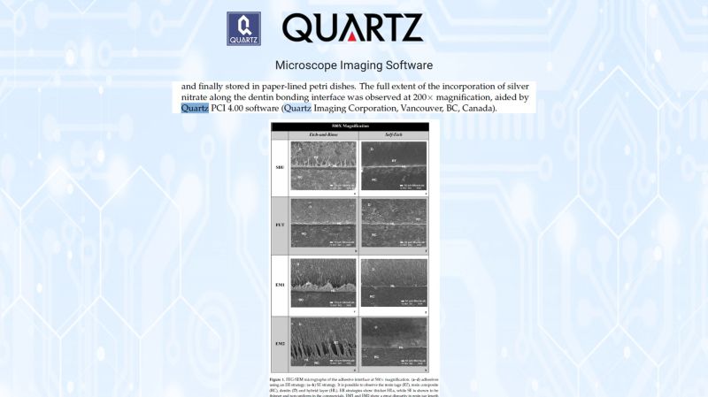
A next-generation dental adhesive has matched commercial standards — without Bisphenol A.
Dental restorations fail from the inside out.
Water seeps into the bond, collagen degrades, and the restoration fails before it should.
Researchers at Instituto Universitário Egas Moniz tackled this by replacing Bis-GMA with a novel dendrimer called G-IEMA, creating a BPA-free adhesive built to last longer.
The obstacle: existing universal adhesives attract and retain water at the bonding interface, the primary driver of long-term failure.
The solution: a reformulated adhesive with lower hydrophilicity, tested against two leading commercial products across four adhesive groups and two bonding strategies.
Quartz PCI software was used to analyze silver nitrate penetration along the dentin interface, delivering the precise imaging needed to quantify nanoleakage at the microscopic level.
After three months of aging, both experimental adhesives showed significantly less nanoleakage than the commercial alternatives.
Bond strength matched commercial adhesives from day one.
The humanity: restorations that hold longer mean fewer repeat procedures, less chair time, and better long-term outcomes for patients.
All rights for the excerpts and images remain with the authors and the full article is found here.
Vasconcelos e Cruz, J., Delgado, A.H.S., Félix, S., Brito, J., Gonçalves, L., & Polido, M. (2022). Improving Properties of an Experimental Universal Adhesive by Adding a Multifunctional Dendrimer (G-IEMA): Bond Strength and Nanoleakage Evaluation. Polymers, 14(7), 1462.
Thank you for mentioning Quartz Imaging in the paper and congratulations on publishing!
Learn more about Quartz Imaging products here.
April 15, 2026
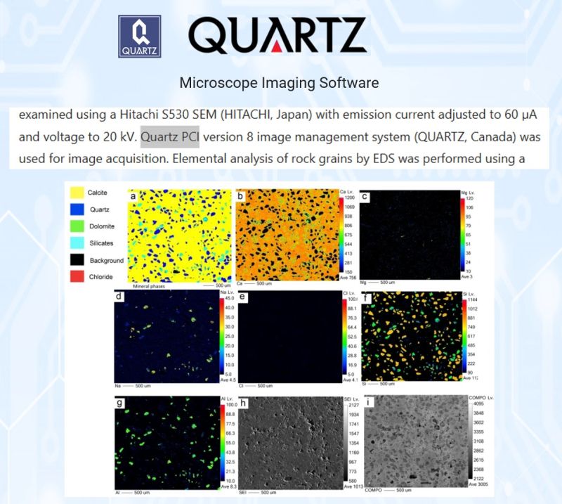
Unlocking a 20-Million-Year-Old Geological Mystery to Better Understand Our Planet
For over 30 years, the origin of unusual dolomite formations in Kuwait's Al-Subiya sabkha had scientists puzzled.
How does a rare mineral form in sediment with no obvious precursor, in the middle of an ancient coastal mud volcano?
That was the obstacle.
A research team from the University of Toronto set out to investigate a recently discovered early-middle Miocene mud volcano outcrop, combining field sampling with geochemical analysis, electron probe microanalysis, and scanning electron microscopy.
Quartz PCI image management software was used for SEM image acquisition, helping the team capture and document the fine-scale mineral structures at the heart of the mystery.
The findings revealed that dolomite formation was driven by an interconnected set of conditions: hydrocarbon seepage, hypersaline seawater, and the burrowing activity of ancient crustaceans that created channels for methane-rich fluids to mix with the surrounding sediment.
Understanding how and where dolomite forms matter beyond geology.
Dolomite-bearing formations are significant hydrocarbon reservoirs worldwide, and this research adds a critical piece to our understanding of how Earth's subsurface systems work.
All rights for the excerpts and images remain with the authors and the full article is found here.
Alibrahim, A., Duane, M. J., & Dittrich, M. (2021). Dolomite genesis in bioturbated marine zones of an early-middle Miocene coastal mud volcano outcrop (Kuwait). Scientific Reports, 11, 6636.
Thank you for mentioning Quartz Imaging in the paper and congratulations on publishing!
Learn more about Quartz Imaging products here.